43 labels of the human brain
Normal chest MDCT with anatomic labels | e-Anatomy - e-Anatomy … 10/03/2022 · Pocket Atlas of Human Anatomy: 5th edition - W. Dauber, Founded by Heinz Fene Anatomical variants and notes from the author about the anatomical labeling of the thorax CT: In the lower lobe of the left lung, there is an inconstant subsuperior pulmonary segment that is seen in approximately 30% of individuals, located between the superior and basal segments of the … Brain Anatomy and How the Brain Works | Johns Hopkins Medicine Gray and white matter are two different regions of the central nervous system. In the brain, gray matter refers to the darker, outer portion, while white matter describes the lighter, inner section underneath. In the spinal cord, this order is reversed: The white matter is on the outside, and the gray matter sits within.
› science-center › techThe Role of Dopamine as a Neurotransmitter in the Human Brain However, between 1957 and 1959, parallel efforts by Kathleen Montagu and her colleagues at Hans Weil-Malherbe lab at Runwell Hospital (England) and Arvin Carlsson and his colleagues at Lund University (Sweden) helped lead to the initial findings that would collectively suggest dopamine’s role as a neurotransmitter in the human brain.

Labels of the human brain
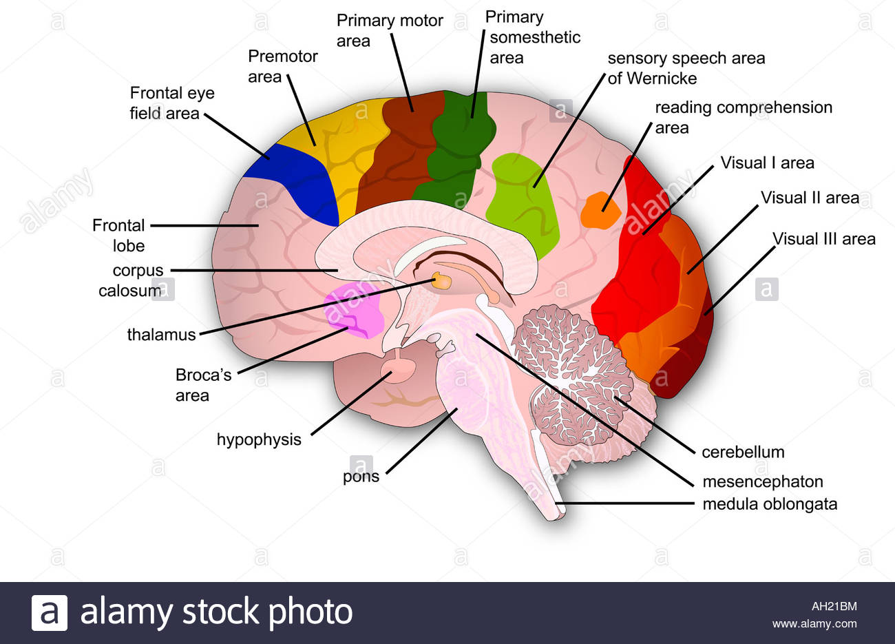
Frontiers | 101 Labeled Brain Images and a Consistent Human Cortical ... We introduce the Mindboggle-101 dataset, the largest and most complete set of free, publicly accessible, manually labeled human brain images. To manually label the macroscopic anatomy in magnetic resonance images of 101 healthy participants, we created a new cortical labeling protocol that relies on robust anatomical landmarks and minimal manual edits after initialization with automated labels ... Labeled Brain Model Diagram | Science Trends The frontal lobe of the brain is responsible for our critical thinking, planning, reasoning, and problem-solving, as well as our experience of emotions. The rear portion of the frontal lobe is the motor cortex, which receives inputs from the other lobes and carries out the movements of the body associated with them. Automated Labeling of the Human Brain - PMC - NCBI There are two, literally opposite, coordinate-based methods to retrieve brain labels from an atlas for a brain image. Both rely on spatial normalization to ...
Labels of the human brain. 1663 Brain diagram with labels Images, Stock Photos & Vectors Human brain vector illustration. Labeled anatomical educational head organ parts scheme separated by colors. The Role of Dopamine as a Neurotransmitter in the Human Brain To understand how dopamine functions in the human brain as a neurotransmitter requires looking at the effect of dopamine binding to D1-like and D2-like receptor types to exert their effects via second messenger systems. The binding of dopamine to D1-like receptors (D1 and D5) results in excitation via the opening of Na+ channels or inhibition via the opening of K+ channels. D1 … automated labeling of neuroanatomical structures in the human brain In contrast to existing segmentation procedures that only label a small number of tissue classes, the current method assigns one of 37 labels to each voxel, ... Human Brain: Facts, Functions & Anatomy | Live Science The human brain weighs about 3 lbs. (1.4 kilograms) and makes up about 2% of a human's body weight. On average, male brains are about 10% larger than female brains, according to Northwestern ...
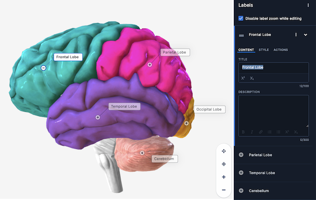
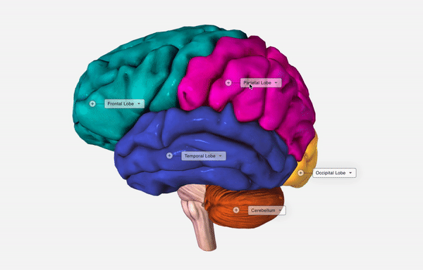
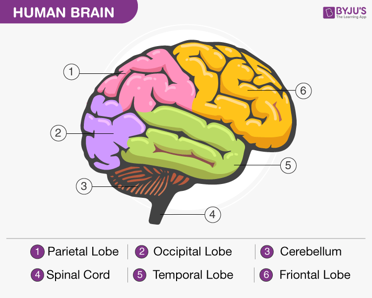
SARS-CoV-2 infection and persistence throughout the human body and brain 03/12/2021 · distribution, replication, and cell-type specificity across the human body, including brain, 73 from acute infection through over seven months following symptom onset. We show that 74 Anatomy of the Brain: Structures and Their Function - ThoughtCo The brain and spinal cord are the two main structures of the central nervous system. There are three major divisions of the brain. They are the forebrain, the midbrain, and the hindbrain. Key Takeaways The forebrain, the midbrain, and the hindbrain are the three main parts of the brain. Amazon.com: XINDAM 3D Human Brain with Labels Anatomical Model ... This item: XINDAM 3D Human Brain with Labels Anatomical Model Paperweight (Laser Etched) in Crystal Glass Ball Science Gift (Included LED Base) $66.99 Brain 11 Ounce Ceramic Coffee Mug (WC462M) $18.98 Anatomic Brain Specimen Coasters (Set of 10) - Neuroscience Gifts, Gifts for Medical Student Gifts Brain Decor Human Anatomy Gifts Human Brain - Structure, Diagram, Parts Of Human Brain - BYJUS The cerebrum is the largest part of the brain. It consists of the cerebral cortex and other subcortical structures. It is composed of two cerebral hemispheres that are joined together by heavy, dense bands of fibre called the corpus callosum. The cerebrum is further divided into four sections or lobes:
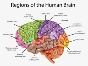
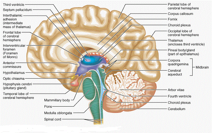
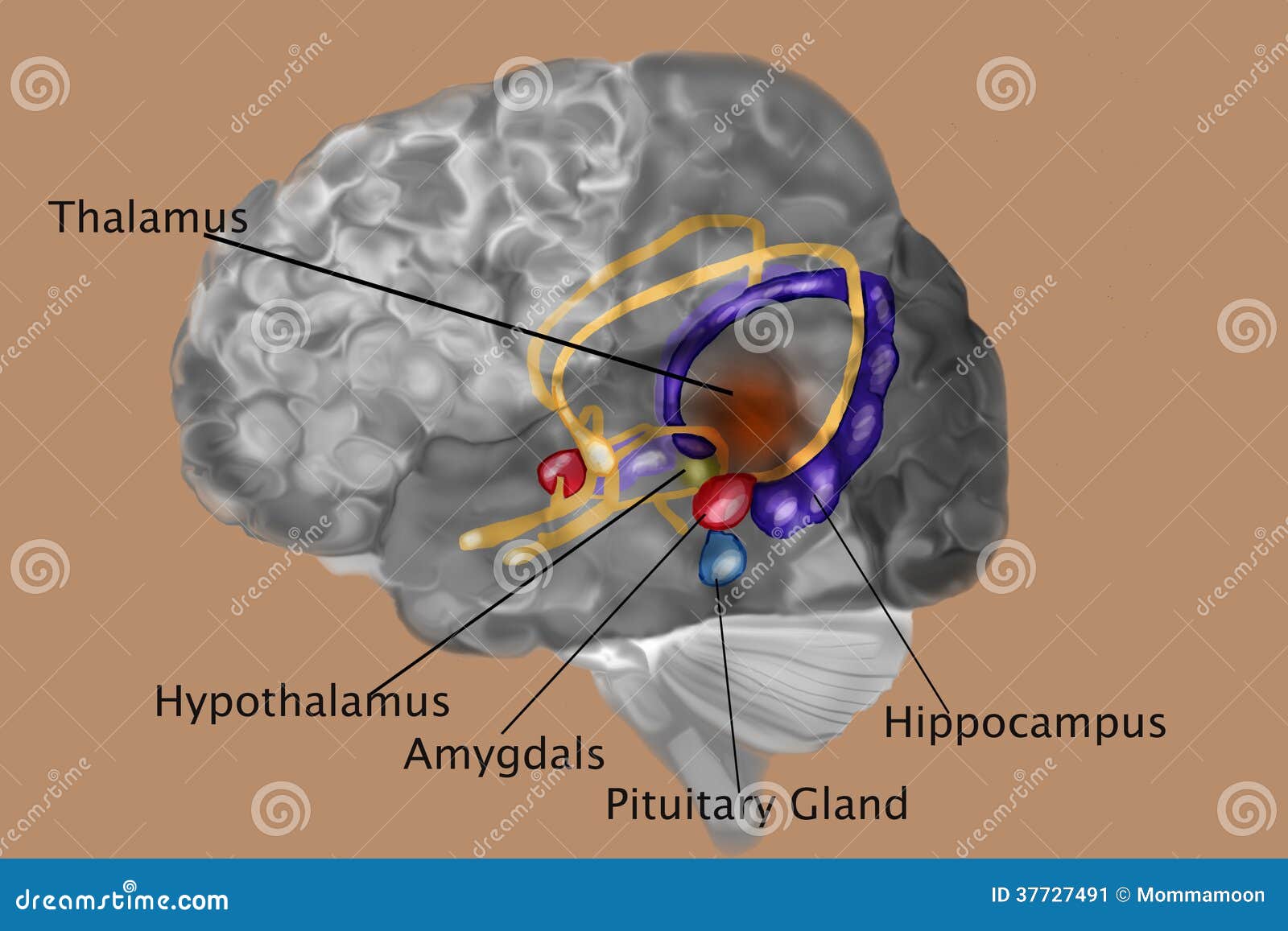
Label the Brain Worksheets (SB11585) - SparkleBox - Pinterest Sep 30, 2018 - A set of diagrams of the brain. ... Label the Brain Worksheets (SB11585) - SparkleBox Human Brain Diagram, Brain Structure Diagram. Labeled Diagrams of the Human Brain You'll Want to Copy Now The central core consists of the thalamus, pons, cerebellum, reticular formation and medulla. These five regions are the central areas that regulate breathing, pulse, arousal, balance, sleep and early stages of processing sensory information. The thalamus interprets the sensory information and helps determine what is good and bad. Labeled Human Brain Illustrations, Royalty-Free Vector ... - iStock Browse 109 labeled human brain stock illustrations and vector graphics available royalty-free, or start a new search to explore more great stock images and vector art. Brain functions vector illustration. Labeled explanation organ... Brain functions vector illustration. Labeled explanation head organ parts scheme. Brain: Atlas of human anatomy with MRI - e-Anatomy - IMAIOS Choroid plexus of fourth ventricle Choroid plexus of lateral ventricle Choroid plexus of third ventricle Choroidal fissure Cingulate gyrus Cingulate sulcus Cingulum Circular sulcus of insula Cistern of lamina terminalis Cistern of lateral cerebral fossa Claustrum Collateral eminence Collateral sulcus
Pin on Bizarro!
Human brain with labels, illustration Stock Photo - Alamy Download this stock image: Human brain with labels, illustration. - PHBYR3 from Alamy's library of millions of high resolution stock photos, ...
Draw a human brain, label the parts and explain the functions ...
Self-Taught AI May Have a Lot in Common With the Human Brain And brains learn from their mistakes on their own, too—only a small part of our brain's feedback comes from an external source saying, essentially, "wrong answer.". The computational ...
How The Brain Works! - Lessons - Blendspace
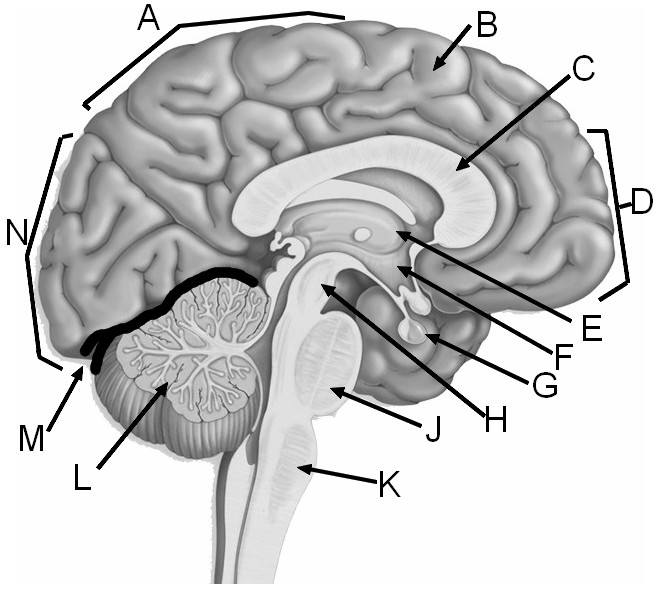
Solved This cross section of the human brain depicts several - Chegg Psychology questions and answers. This cross section of the human brain depicts several key structures. Of the choices provided, which correctly labels the structures in the drawing? 5 points Save Answ 1 - hypothalamus, 2 nucleus, 3 axon, A4-myelin sheath 1 - hippocampus, 2-reticular formation, 3.1 corpus callosum, 2-, 3-cerebellum 4 ...

Amazon.com : Human Brain Model with Labels, Human Brain Model ...
Parts of the brain: Learn with diagrams and quizzes | Kenhub Labeled brain diagram First up, have a look at the labeled brain structures on the image below. Try to memorize the name and location of each structure, then proceed to test yourself with the blank brain diagram provided below. Labeled diagram showing the main parts of the brain Blank brain diagram (free download!)

Brain Meninges Layers With Labels High-Res Vector Graphic ...
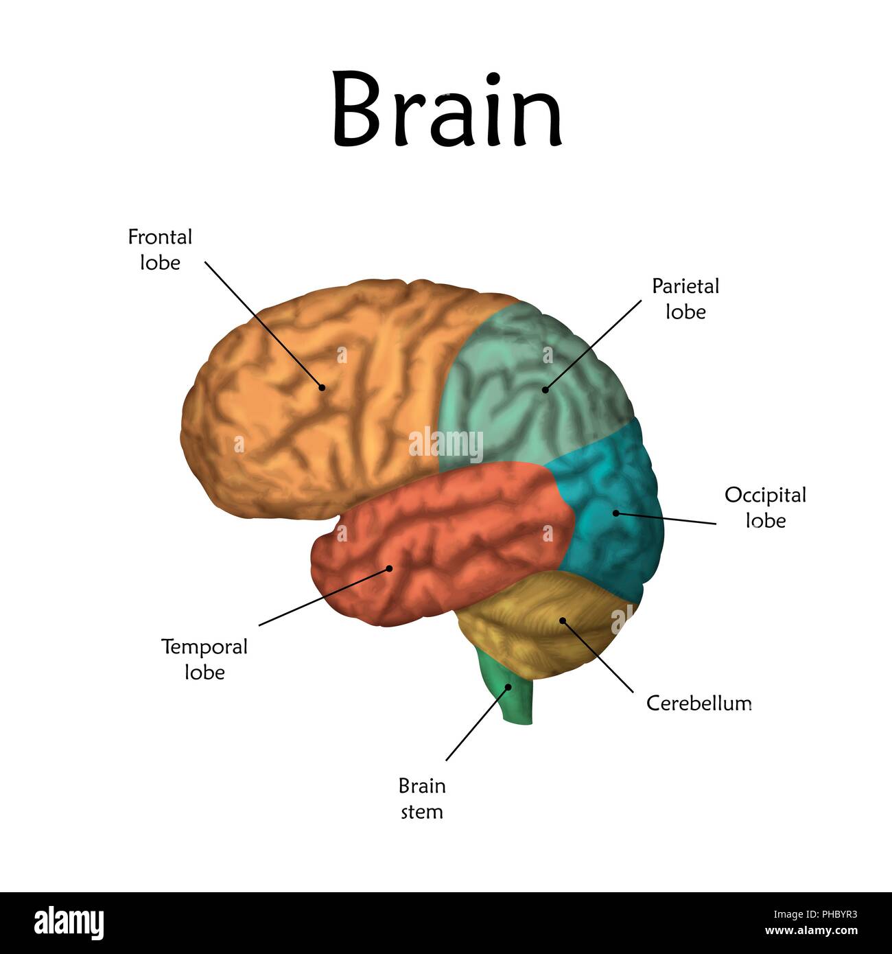
Brain: Anatomy, Pictures, Functions, and Conditions - Verywell Mind The cerebral cortex is the part of the brain that makes human beings unique. Functions that originate in the cerebral cortex include: Consciousness Higher-order thinking Imagination Information processing Language Memory Perception Reasoning Sensation Voluntary physical action 1 The cerebral cortex is what we see when we look at the brain.
Edit a label in my model – Human Support
3D Brain This interactive brain model is powered by the Wellcome Trust and developed by Matt Wimsatt and Jack Simpson; reviewed by John Morrison, Patrick Hof, and Edward Lein. Structure descriptions were written by Levi Gadye and Alexis Wnuk and Jane Roskams .

Buy BEAMNOVA Human Brain Model 2 Times Life Size for ...
Labeled Brain Images – Browse 734 Stock Photos, Vectors, and Video Colored and labeled human brain diagram. Human Brain Sagittal Section with Labels. Human brain anatomy, labeled. Anatomy of the human brain.Sagittal cut.

Modern Digital Illustration Human Brain Stock Photo by ...
The Human Brain - Visible Body The brain gives us self-awareness and the ability to speak and move in the world. Its four major regions make this possible: The cerebrum, with its cerebral cortex, gives us conscious control of our actions. The diencephalon mediates sensations, manages emotions, and commands whole internal systems. The cerebellum adjusts body movements, speech ...
Brain – Human Brain Diagrams and Detailed Information
Human Brain Diagram - Labeled, Unlabled, and Blank The composition of the mind especially 7 Diagram Of The Human Brain is complex due its complex framework and also feature. This incredible body organ functions as a control center by getting, analyzing, and also driving physical info throughout the physical body. #humanbrain #humanbrainpicture #brainpicture
Human Brain No Labels - Illustration Transparent PNG ...
2,831 Labeled brain anatomy Images, Stock Photos & Vectors - Shutterstock 2,831 labeled brain anatomy stock photos, vectors, and illustrations are available royalty-free. See labeled brain anatomy stock video clips Image type Orientation Artists Sort by Popular Healthcare and Medical Anatomy human brain brain organ cerebellum medicine human body cerebrum cerebral cortex Next of 29
Label the Brain Worksheets (SB11585) - SparkleBox
Whole Brain Segmentation: Automated Labeling of ... Jan 31, 2002 ... Typically, manual labeling of brain structures is accomplished using a variety of information including image intensities, global position ...
Brain Surface Anatomy, With Labels Art Print by Alan Gesek | iCanvas
e-Anatomy: radiologic anatomy atlas of the human body - e-Anatomy - IMAIOS e-Anatomy is an award-winning interactive atlas of human anatomy. It is the most complete reference of human anatomy available on web, iPad, iPhone and android devices. Explore over 6,700 anatomic structures and more than 870,000 translated medical labels. Images in: CT, MRI, Radiographs, Anatomic diagrams and nuclear images. Available in 12 ...
Brain Label
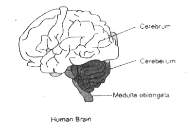
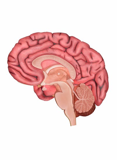
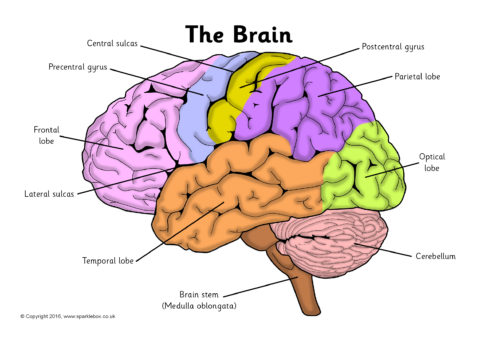
Human brain - Wikipedia The brainstem includes the midbrain, the pons, and the medulla oblongata. Behind the brainstem is the cerebellum ( Latin: little brain ). [8] The cerebrum, brainstem, cerebellum, and spinal cord are covered by three membranes called meninges. The membranes are the tough dura mater; the middle arachnoid mater and the more delicate inner pia mater.

Draw the diagram of human brain. Explain its functions.
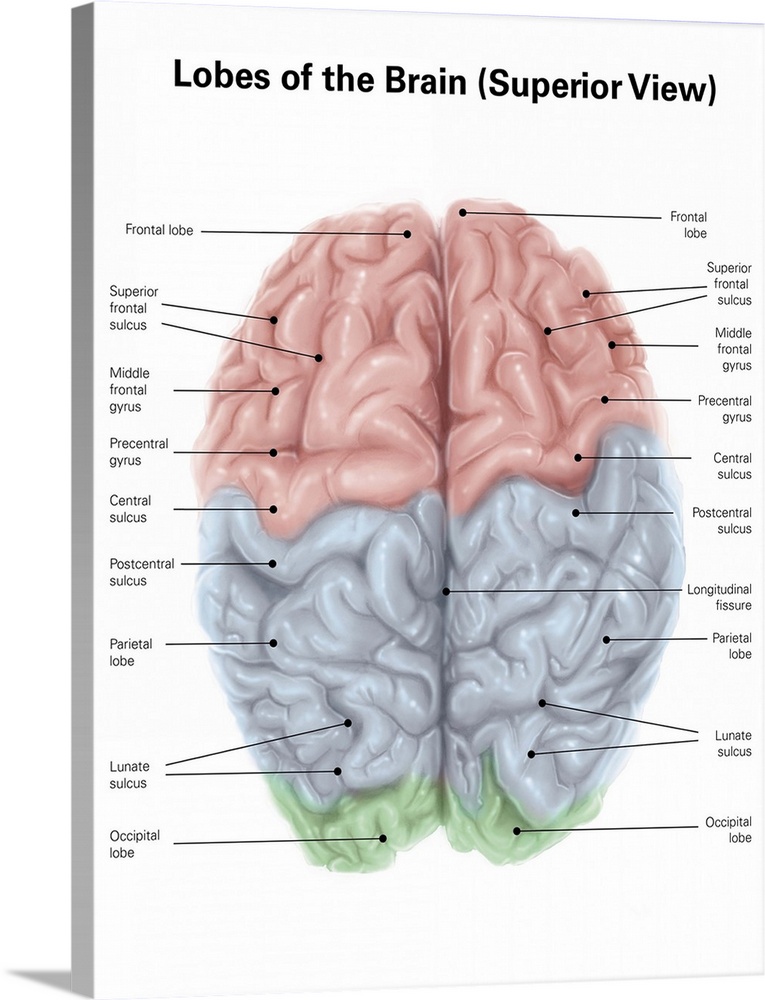
Diagram Of Brain with their Labelings and Detailed Explanation - BYJUS The parietal lobe is found at the upper back of our brain. This lobe functions by controlling all our complex behaviours, including senses of vision, the sense of touch, spatial orientation and body awareness. It manages body position, movements, the perception of stimuli, orientation, handwriting and visuospatial processing. The Occipital Lobe

Sagittal section of the human brain with cerebrum and ...
› en › e-Anatomye-Anatomy: radiologic anatomy atlas of the human body - IMAIOS e-Anatomy is an award-winning interactive atlas of human anatomy. It is the most complete reference of human anatomy available on web, iPad, iPhone and android devices. Explore over 6,700 anatomic structures and more than 870,000 translated medical labels. Images in: CT, MRI, Radiographs, Anatomic diagrams and nuclear images. Available in 12 ...
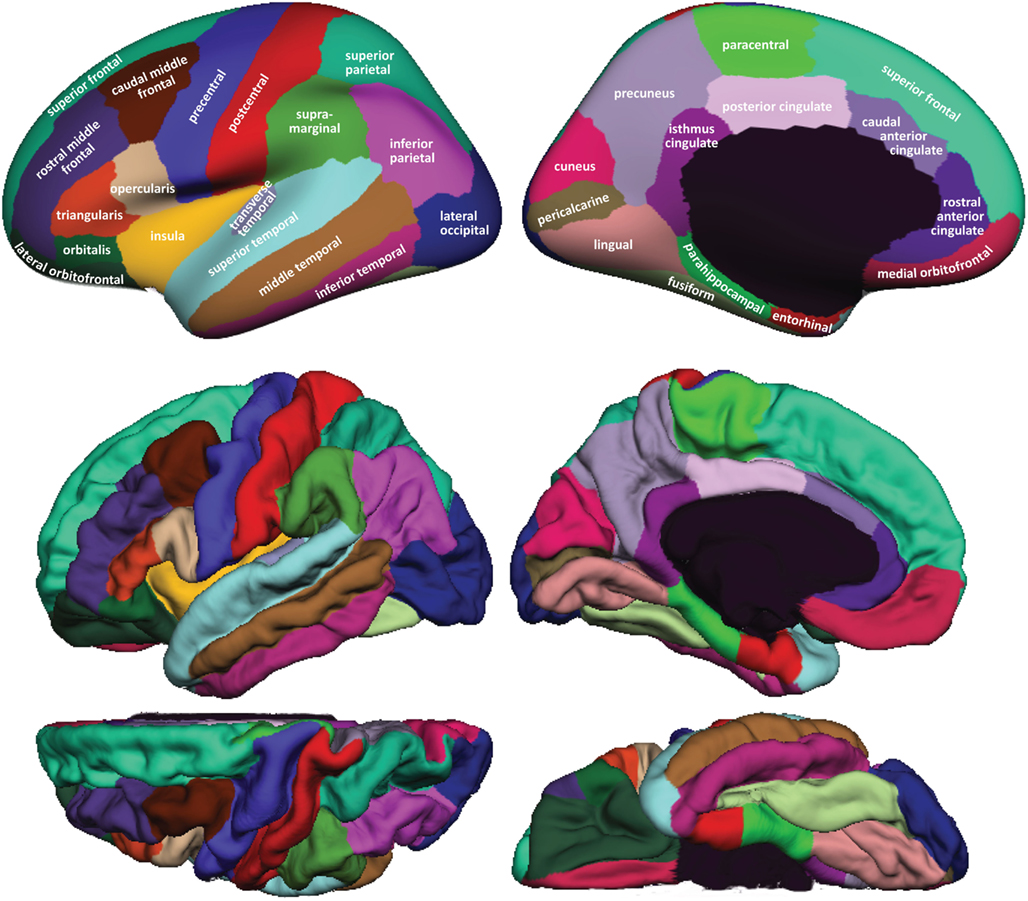
Frontiers | 101 Labeled Brain Images and a Consistent Human ...
Brain - Human Brain Diagrams and Detailed Information - Innerbody The brainstem is made of three regions: the medulla oblongata, the pons, and the midbrain. A net-like structure of mixed gray and white matter known as the reticular formation is found in all three regions of the brainstem. The reticular formation controls muscle tone in the body and acts as the switch between consciousness and sleep in the brain.
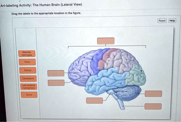
SOLVED: Art-labeling Activity: The Human Brain (Lateral View ...
humanstress.caHome - CESH / CSHS The Centre for Studies on Human Stress (CSHS) is dedicated to improving the physical and mental health of Canadians by empowering individuals with scientifically grounded information on the effects of stress on the brain and body.

AP label Human brain Flashcards | Quizlet
How to draw and label human brain step by step.(full tutorial) Jun 25, 2018 ... hello friends in this video I have shown you how to draw human mind and label it step by step. friends I have explain all the parts of the ...

How to draw human brain/ draw labelled diagram of brain/brain diagram/draw and label brain diagram
Labeled Parts Of The Brain Illustrations, Royalty-Free Vector ... - iStock detailed anatomy of the human brain. detailed anatomy of the human brain. Illustration showing the medulla, pons, cerebellum, hypothalamus, thalamus, midbrain. Sagittal view of the brain. Isolated on a white background. Pineal gland anatomical cross section vector illustration...

Label the Brain Worksheets (SB11585) - SparkleBox | Human ...
Nervous System - Label the Brain - TheInspiredInstructor.com Nervous System - Label the Brain Nervous System - Brain Name: Choose the correct names for the parts of the brain. ( 1) (2) (3) (4) (5) (6) (7) (8) ( 9) This brain part controls thinking. (10) This brain part controls balance, movement, and coordination. (11) This brain part controls involuntary actions such as breathing, heartbeats, and digestion.
draw neat diagram of human brain and label medula and ...
Home - CESH / CSHS The Centre for Studies on Human Stress (CSHS) is dedicated to improving the physical and mental health of Canadians by empowering individuals with scientifically grounded information on the effects of stress on the brain and body. Search here: Home; Our Impact; Become a member; Glossary; Contact; Facebook; Français; Stress Stress. WHAT IS STRESS? History of stress; …
Solved] Label the parts of the human brain and describe their ...
› en › e-AnatomyNormal chest MDCT with anatomic labels | e-Anatomy - IMAIOS Mar 10, 2022 · Pocket Atlas of Human Anatomy: 5th edition - W. Dauber, Founded by Heinz Fene Anatomical variants and notes from the author about the anatomical labeling of the thorax CT: In the lower lobe of the left lung, there is an inconstant subsuperior pulmonary segment that is seen in approximately 30% of individuals, located between the superior and ...
Superior View Of Human Brain With Colo - Canvas Art Print | Alan Gesek
en.wikipedia.org › wiki › File:Human_skeleton_frontFile:Human skeleton front en.svg - Wikipedia Restructured the image internals by adding layers, changing groupings, and adding meaningful ids and labels so that the image is easier to manipulate programmatically. Also made the labels text elements and gave them ids (it might be possible to generate
Vevor Human Brain Model Anatomy 4-part Model Of Brain W/labels & Display Base Color-coded Life Size Science Classroom Display - Medical Science - ...
File:Human skeleton front en.svg - Wikipedia English: diagram of a human female skeleton. the Red lines point individual bones and the names are writen in singular, the blue lines connect to group of bones and are in plural form. Date : 3 January 2007: Source: Own work. Image renamed from File:Human skeleton front.svg: Author: LadyofHats Mariana Ruiz Villarreal: Permission (Reusing this file) Public domain Public domain …
Human brain with labels, illustration Stock Photo - Alamy
The human brain: Parts, function, diagram, and more - Medical News Today It is made up of three major areas: the cerebrum, cerebellum, and brain stem. It controls critical biological processes that are crucial for survival, such as breathing and temperature regulation....
Human Brain with Labels stock illustration. Illustration of ...
Brain (Human Anatomy): Picture, Function, Parts, Conditions ... - WebMD • The cortex is the outermost layer of brain cells. Thinking and voluntary movements begin in the cortex. • The brain stem is between the spinal cord and the rest of the brain. Basic functions like...

brain diagram labeled | Brain diagram, Brain stem, Corpus ...
The Human Brain Atlas - Michigan State University In this atlas you can view MRI sections through a living human brain as well as corresponding sections stained for cell bodies or for nerve fibers. The stained sections are from a different brain than the one which was scanned for the MRI images. Furthermore, for the stained sections, the brain was removed from the skull, dehydrated, embedded ...

Cerebrum - Wikipedia
› tech › 2020/01/22Harvard Embraces Debunked 'Implicit Bias' Test that Labels ... Jan 22, 2020 · A popular quiz on Harvard University's website was designed in 1998 by psychologists to determine a person's level of subconscious racism. Although the test has been thoroughly debunked by researchers since its introduction, it has remained a fixture of progressive activism.

Label the parts of human brain.Bс - Brainly.in
human brain with labels human brain with labels human brain with labels Brain superior human colored lobes labels alamy. Occipital bone of the human skull. Anatomy cerebral venous brain mri vascular scan system sinus thrombosis thebluntdissection venogram related cerebri pseudotumor human brain with labels

ventral aspect of human brain labeling Diagram | Quizlet
The Human Brain | Brain and Cognitive Sciences | MIT OpenCourseWare This course surveys the core perceptual and cognitive abilities of the human mind and asks how they are implemented in the brain. Key themes include the representations, development, and degree of functional specificity of these components of mind and brain. The course will take students straight to the cutting edge of the field, empowering them to understand and critically evaluate empirical ...
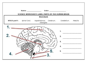
Science worksheets: Label parts of a human brain by Science ...
101 Labeled Brain Images and a Consistent Human Cortical Labeling ... Labeling the macroscopic anatomy of the human brain is instrumental in educating biologists and clinicians, visualizing biomedical data, localizing brain data for identification and comparison, and perhaps most importantly, subdividing brain data for analysis.

Sagital Section Of The Human Brain With Regions And Labels Wall Peel
Amazon.com: brain model labeled Generies 2021 Newest Design Human Skull Anatomical Model,with Painted Sutures 54 Pcs Labeled Numbered Skull Models for Medical Students,Human Brain Model for Kids Drawing Anatomy 64 $3250 FREE delivery Mon, Oct 10 Only 18 left in stock - order soon.

Vector Isolated Illustration Of Brain Components In Man Head ...
› publication › 357197928_SARSSARS-CoV-2 infection and persistence throughout the human ... Dec 03, 2021 · of the brain was accomplished in 11 of the 44 cases (Fig. 1). The cohort was 29.5% female with 127 a mean age of 59.2 years and was diverse across race and ethnicity (Extended Data Table 1).

3D Human Brain with Labels Anatomical Model Statue Paperweight(Laser Etched) in Crystal Glass Cube Science Gift (No Included LED Base)(2.3x2.3x2.3 ...
Automated Labeling of the Human Brain - PMC - NCBI There are two, literally opposite, coordinate-based methods to retrieve brain labels from an atlas for a brain image. Both rely on spatial normalization to ...
Superior view of human brain with colored lobes and labels Solid-Faced Canvas Print
Labeled Brain Model Diagram | Science Trends The frontal lobe of the brain is responsible for our critical thinking, planning, reasoning, and problem-solving, as well as our experience of emotions. The rear portion of the frontal lobe is the motor cortex, which receives inputs from the other lobes and carries out the movements of the body associated with them.
Draw and label the diagrams: Human brain. - Sarthaks eConnect ...
Frontiers | 101 Labeled Brain Images and a Consistent Human Cortical ... We introduce the Mindboggle-101 dataset, the largest and most complete set of free, publicly accessible, manually labeled human brain images. To manually label the macroscopic anatomy in magnetic resonance images of 101 healthy participants, we created a new cortical labeling protocol that relies on robust anatomical landmarks and minimal manual edits after initialization with automated labels ...
How do labels work in the BioDigital Human? – Human Support

How to draw human brain/draw and label brain diagram/draw labelled diagram of brain/brain diagram

Human Brain Anatomy Lateral View With Labels High-Res Vector ...

Human Brain Diagram Template - SlideModel
Human Brain - Structure, Diagram, Parts Of Human Brain
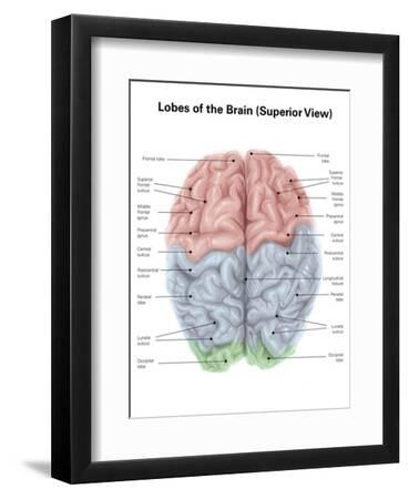
Superior View of Human Brain with Colored Lobes and Labels ...
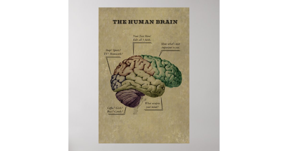
Customizable Human Brain Poster, edit the 5 labels Poster | Zazzle
Post a Comment for "43 labels of the human brain"