41 brain mri with labels
A multi-atlas label fusion tool for neonatal brain MRI parcellation and ... Introduction. Structure-by-structure analysis, in which the brain is parcellated based on structural units that follow standard ontology in brain anatomy, is widely used to investigate disease-related changes seen on brain MRI scans. 1-4 Numerous tools for brain parcellation methods have been proposed in the past and their accuracy has continuously improved, especially in the past decade ... en.wikipedia.org › wiki › Diffusion_MRIDiffusion MRI - Wikipedia Diffusion-weighted magnetic resonance imaging (DWI or DW-MRI) is the use of specific MRI sequences as well as software that generates images from the resulting data that uses the diffusion of water molecules to generate contrast in MR images.
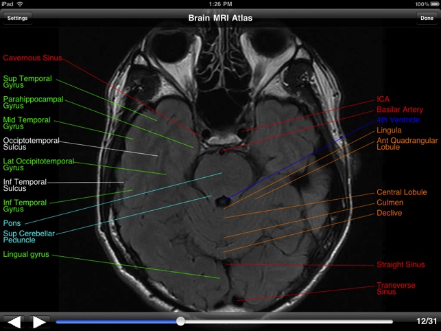
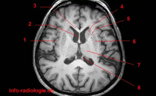
MRI anatomy | free MRI axial brain anatomy - Mrimaster.com This MRI brain cross sectional anatomy tool is absolutely free to use. Use the mouse scroll wheel to move the images up and down alternatively use the tiny arrows (>>) on both side of the image to move the images.
Brain mri with labels
Atlas of BRAIN MRI - W-Radiology Brain magnetic resonance imaging (MRI) is a common medical imaging method that allows clinicians to examine the brain's anatomy (1). It uses a magnetic field and radio waves to produce detailed images of the brain and the brainstem to detect various conditions (2). › health-news › is-it-safe-toIs It Safe to Undergo Multiple MRI Exams? - Healthline Sep 27, 2018 · The findings, at the very least, are a cause for concern. That’s what Dr. Emanuel Kanal says about the Food and Drug Administration’s safety announcement last week on the risk of brain ... Labeled MRI Brain Scans - Neuromorphometrics We can also label scans that you provide and we are very interested in labeling white matter anatomy as seen in diffusion-weighted MRI scans. If you want an aggregate version of our data, we can provide it as a probabilistic atlas. The cost to label a single scan is $2449 (USD).
Brain mri with labels. Labels · sunreef/brain-mri · GitHub A Machine Learning project to determine the age of a patient from the results of an MRI. - Labels · sunreef/brain-mri MRI brain (summary) | Radiology Reference Article - Radiopaedia MRI brain is a specialist investigation that is used for the assessment of a number of neurological conditions. It is the main method to investigate conditions such as multiple sclerosis and headaches, and used to characterize strokes and space-occupying lesions. Reference article MRI Brain Atlas - University of Minnesota This web app Atlas is intended for veterinary students and radiologists seeking quick access to canine brain anatomy through a mobile device. Via a toggle button, either MRI images or approximately comparable Brain Transection images may be viewed with or without labels. Navigation & Labels. › en › e-AnatomyShoulder: MRI, radiographical, and illustrated anatomical ... Sep 13, 2021 · MRI of the shoulder : muscles of the rotator cuff labeled on a sagittal MR slice. An MRI of the shoulder of a healthy subject was performed in the 3 planes of space (coronal, axial, sagittal) commonly used in osteoarticular imaging, with two weightings to explore the musculoskeletal pathology of the shoulder: spin-echo T1 and proton-density ...
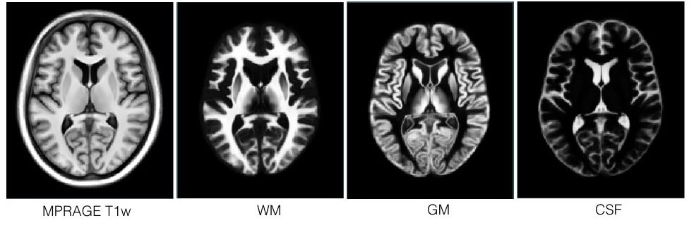
en.wikipedia.org › wiki › Molecular_imagingMolecular imaging - Wikipedia MRI has the advantages of having very high spatial resolution and is very adept at morphological imaging and functional imaging. MRI does have several disadvantages though. First, MRI has a sensitivity of around 10 −3 mol/L to 10 −5 mol/L which, compared to other types of imaging, can be very limiting. This problem stems from the fact that ... Brain MRI: How to read MRI brain scan | Kenhub MRI is the most sensitive imaging method when it comes to examining the structure of the brain and spinal cord. It works by exciting the tissue hydrogen protons, which in turn emit electromagnetic signals back to the MRI machine. The MRI machine detects their intensity and translates it into a gray-scale MRI image. Deep learning from MRI-derived labels enables automatic brain tissue ... Deep learning from MRI-derived labels enables automatic brain tissue classification on human brain CT Neuroimage. 2021 Dec 1;244:118606. doi: 10.1016/j ... Our proposed model predicted brain tissue classes accurately from unseen CT images (Dice coefficients of 0.79, 0.82, 0.75, 0.93 and 0.98 for GM, WM, CSF, brain volume and ICV, respectively). ... MRI head sagittal T1 - labeling questions | Radiology Case ... The labeled structures are (excluding the correct side): temporal horn of lateral ventricle primary fissure of cerebellum choroid plexus trigone (atrium) of lateral ventricle horizontal fissure of cerebellum occipital horn of lateral ventricle intraorbital segment of optic nerve diploic space of parietal bone body of caudate nucleus maxillary sinus
101 Labeled Brain Images and a Consistent Human Cortical Labeling ... Labeled anatomical subdivisions of the brain enable one to quantify and report brain imaging data within brain regions, which is routinely done for functional, diffusion, and structural magnetic resonance images (f/d/MRI) and positron emission tomography data. MRI head axial T2 - labeling questions - Radiopaedia The labeled structures are (excluding the correct side): cervical spinal cord posterior arch of C1 odontoid process (peg or dens) of C2 parotid gland intradural segment (V4) of dominant vertebral artery cisterna magna intradural segment (V4) of non-dominant vertebral artery cerebellar tonsil occipital condyle medulla oblongata brain anatomy | MRI coronal brain anatomy | free MRI cross sectional ... ELBOW AXIAL. WRIST AXIAL. WRIST CORONAL. KNEE CORONAL. KNEE SAGITTAL. ARTERIES UPPER LEG. ARTERIES LOWER LEG. This MRI brain coronal cross sectional anatomy tool is absolutely free to use. Use the mouse scroll wheel to move the images up and down alternatively use the tiny arrows (>>) on both side of the image to move the images. › AANLIB › casesHarvard University Show labels Show list All modalities to: MR-T1 MR-T2 FDG T1/FDG T2/FDG
CaseStacks.com - MRI Brain Anatomy
Choreography Controlled (ChoCo) brain MRI artifact generation for ... Up to our best knowledge, no protocol for generating labeled datasets of MRI images corrupted by controlled motion has yet been proposed. Hence, we present a methodology allowing the acquisition of reproducible motion-corrupted MRI images as well as validation of the system's performance by motion estimation through rigid-body volume ...
Brain: Atlas of human anatomy with MRI - e-Anatomy
Labeled imaging anatomy cases | Radiology Reference Article ... This article lists a series of labeled imaging anatomy cases by body region and modality. Brain CT head: non-contrast axial CT head: non-contrast coronal CT head: non-contrast sagittal CT head: angiogram axial CT head: angiogram coronal CT...

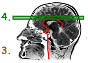
Normal anatomy of the brain on sagittal plane T1weighted ...
Cross-sectional anatomy of the brain - e-Anatomy - IMAIOS An MRI was performed on a healthy subject, with several acquisitions with different weightings: spin-echo T1, T2 and FLAIR, T2 gradient-echo, diffusion, and T1 after gadolinium injection. We obtained 24 axial slices of the normal brain.

NeuroRad for iPad is a great app for medical professionals to ...
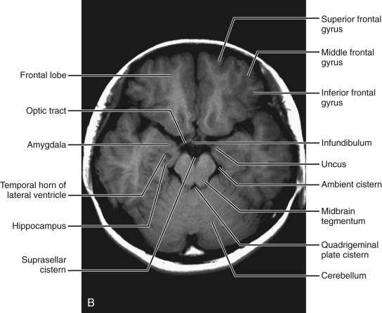
Brain: Atlas of human anatomy with MRI - e-Anatomy - IMAIOS MRI Atlas of the Brain. This page presents a comprehensive series of labeled axial, sagittal and coronal images from a normal human brain magnetic resonance imaging exam. This MRI brain cross-sectional anatomy tool serves as a reference atlas to guide radiologists and researchers in the accurate identification of the brain structures.
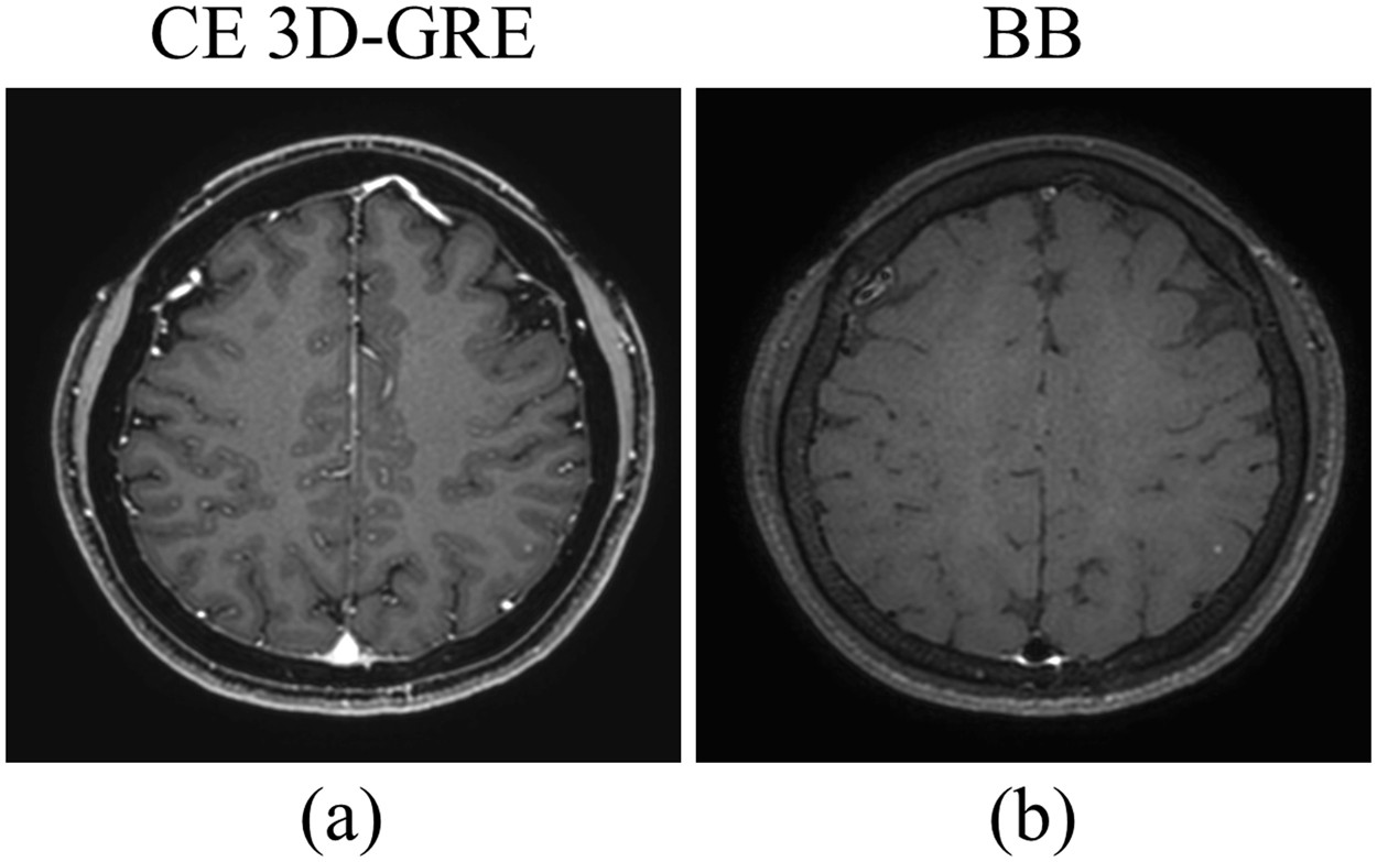
Deep-learned 3D black-blood imaging using automatic labelling ...
› en › e-AnatomyNormal chest MDCT with anatomic labels | e-Anatomy - IMAIOS Mar 10, 2022 · IMAIOS and selected third parties, use cookies or similar technologies, in particular for audience measurement. Cookies allow us to analyze and store information such as the characteristics of your device as well as certain personal data (e.g., IP addresses, navigation, usage or geolocation data, unique identifiers).
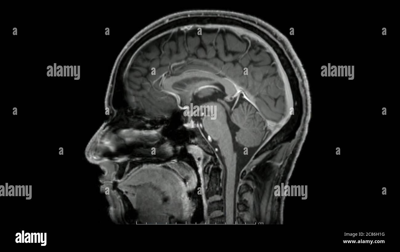
Magnetic resonance images of the brain (MRI brain) sagittal ...
Brain MRI: What It Is, Purpose, Procedure & Results - Cleveland Clinic A brain MRI (magnetic resonance imaging) scan, also called a head MRI, is a painless procedure that produces very clear images of the structures inside of your head — mainly, your brain. MRI uses a large magnet, radio waves and a computer to produce these detailed images. It doesn't use radiation.

How much does a brain MRI cost? | From $225
Labels · alessandrolamberti/brain-mri-segmentation · GitHub Brain MRI segmentation with Resnext50 as backbone - A Pytorch implementation - Labels · alessandrolamberti/brain-mri-segmentation

volBrain: Automated MRI Brain volumetry system
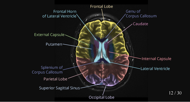
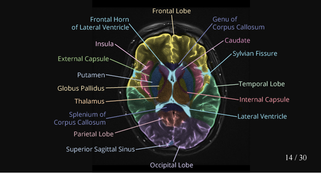
Brain lobes - annotated MRI | Radiology Case | Radiopaedia.org Debowski, M. Brain lobes - annotated MRI. Case study, Radiopaedia.org. (accessed on 30 Sep 2022)

Neuroimaging: Visualize 3D MRI Brain Scans with Python
medicaldecathlon.comMedical Segmentation Decathlon Brain Tumours Target: Gliomas segmentation necrotic/active tumour and oedema Modality: Multimodal multisite MRI data (FLAIR, T1w, T1gd,T2w) Size: 750 4D volumes (484 Training + 266 Testing) Source: BRATS 2016 and 2017 datasets. Challenge: Complex and heterogeneously-located targets

Neural Structure Quiz
Researchers automate brain MRI image labeling, more than ... - ScienceDaily Researchers have automated brain MRI image labeling, needed to teach machine learning image recognition models, by deriving important labels from radiology reports and accurately assigning them to...

volBrain: Automated MRI Brain volumetry system
Labeled MRI Brain Scans - Neuromorphometrics We can also label scans that you provide and we are very interested in labeling white matter anatomy as seen in diffusion-weighted MRI scans. If you want an aggregate version of our data, we can provide it as a probabilistic atlas. The cost to label a single scan is $2449 (USD).

MRI Scans Show The Horrific Effect Cocaine Abuse Can Have On ...
› health-news › is-it-safe-toIs It Safe to Undergo Multiple MRI Exams? - Healthline Sep 27, 2018 · The findings, at the very least, are a cause for concern. That’s what Dr. Emanuel Kanal says about the Food and Drug Administration’s safety announcement last week on the risk of brain ...
Brain Lesion Detection in MRI Images with Graph-cut Algorithms
Atlas of BRAIN MRI - W-Radiology Brain magnetic resonance imaging (MRI) is a common medical imaging method that allows clinicians to examine the brain's anatomy (1). It uses a magnetic field and radio waves to produce detailed images of the brain and the brainstem to detect various conditions (2).
fMRI: Arterial Spin Labeling
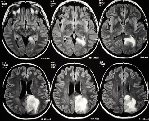
Unusual Brain MRI Findings in Patients Imaged for Headache: a ...

Diagnostic usefulness of arterial spin labeling in MR ...
Brain MRI Atlas on the App Store

7.0 T MRI Axial Images. (a) An axial view image obtained by ...
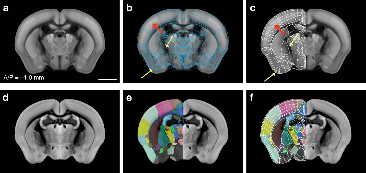
Enhanced and unified anatomical labeling for a common mouse ...

Cross-sectional anatomy of the brain - e-Anatomy
Multi-contrast PD25 atlas – NIST

Approach to MRI brain | LearningNeurology.com
CaseStacks.com - MRI Brain Anatomy

MRI anatomy | free MRI axial brain anatomy
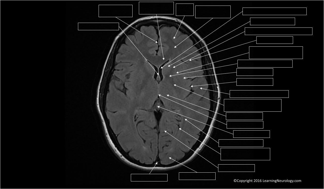
Approach to MRI brain | LearningNeurology.com

Early postmortem brain MRI findings in COVID-19 non-survivors ...

Intelligent Scanning Using Deep Learning for MRI — The ...
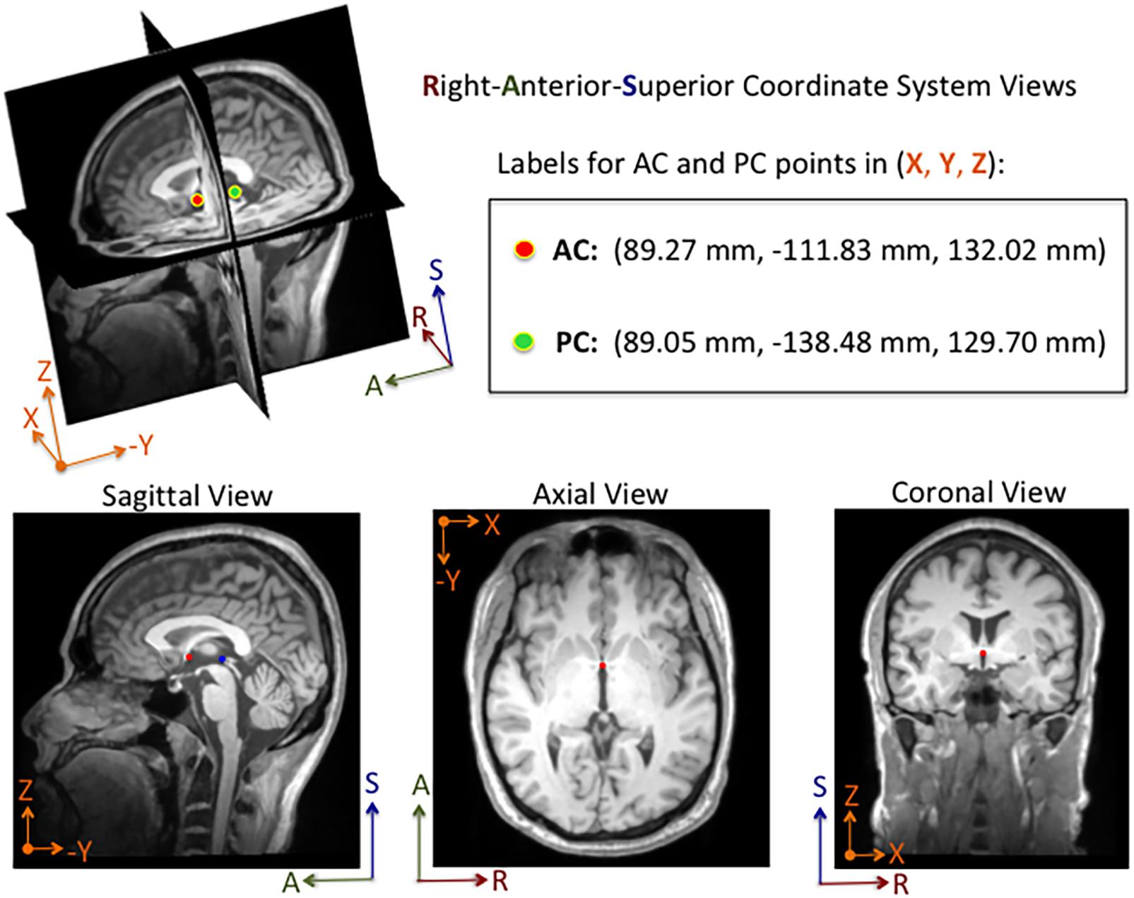
Frontiers | DeepNavNet: Automated Landmark Localization for ...
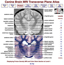
Veterinary Planar Anatomy Courseware
Atlas of BRAIN MRI - W-Radiology

Brain Anatomy and Images Brain

Magnetic Resonance Imaging (MRI): Brain - Connecticut Children's

Tips and traps in brain MRI: Applications to vascular ...

Brain lobes - annotated MRI | Radiology Case | Radiopaedia.org
Normal Anatomy | Radiology Key

MRI anatomy | free MRI axial brain anatomy
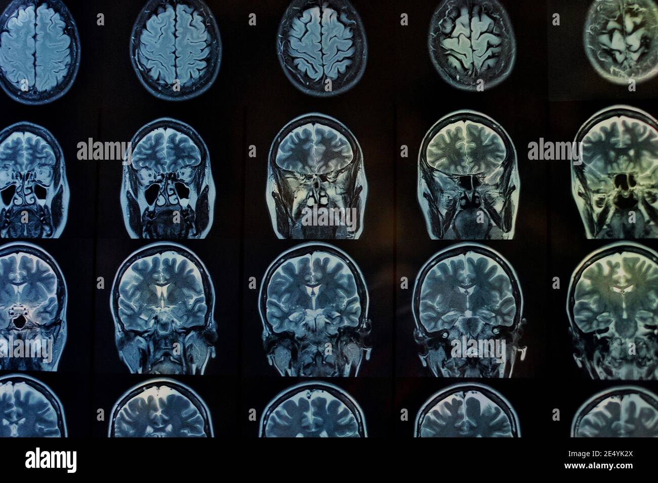
Brain scan mri hi-res stock photography and images - Alamy
Brain MRI: How to read MRI brain scan | Kenhub

Brain Anatomy and Images Brain

MRI anatomy | free MRI axial brain anatomy

MRI anatomy | free MRI axial brain anatomy
Post a Comment for "41 brain mri with labels"