45 diagram of a human cell with labels
Human Anatomy Label Me! Printouts - EnchantedLearning.com Human Anatomy Label Me! Elementary-level Printouts. Read the definitions then label the diagrams. Advertisement. EnchantedLearning.com is a user-supported site. As a bonus, site members have access to a banner-ad-free version of the site, with print-friendly pages. ... Animal Cell Anatomy Label the animal cell diagram using the glossary of ... Skeletal System - Labeled Diagrams of the Human Skeleton The skeletal system's cell matrix acts as our calcium bank by storing and releasing calcium ions into the blood as needed. Proper levels of calcium ions in the blood are essential to the proper function of the nervous and muscular systems. Bone cells also release osteocalcin, a hormone that helps regulate blood sugar and fat deposition.
Labeled Diagram of the Human Kidney - Bodytomy Labeled Diagram of the Human Kidney The human kidneys house millions of tiny filtration units called nephrons, which enable our body to retain the vital nutrients, and excrete the unwanted or excess molecules as well as metabolic wastes from the body. In addition, they also play an important role in maintaining the water balance of our body.
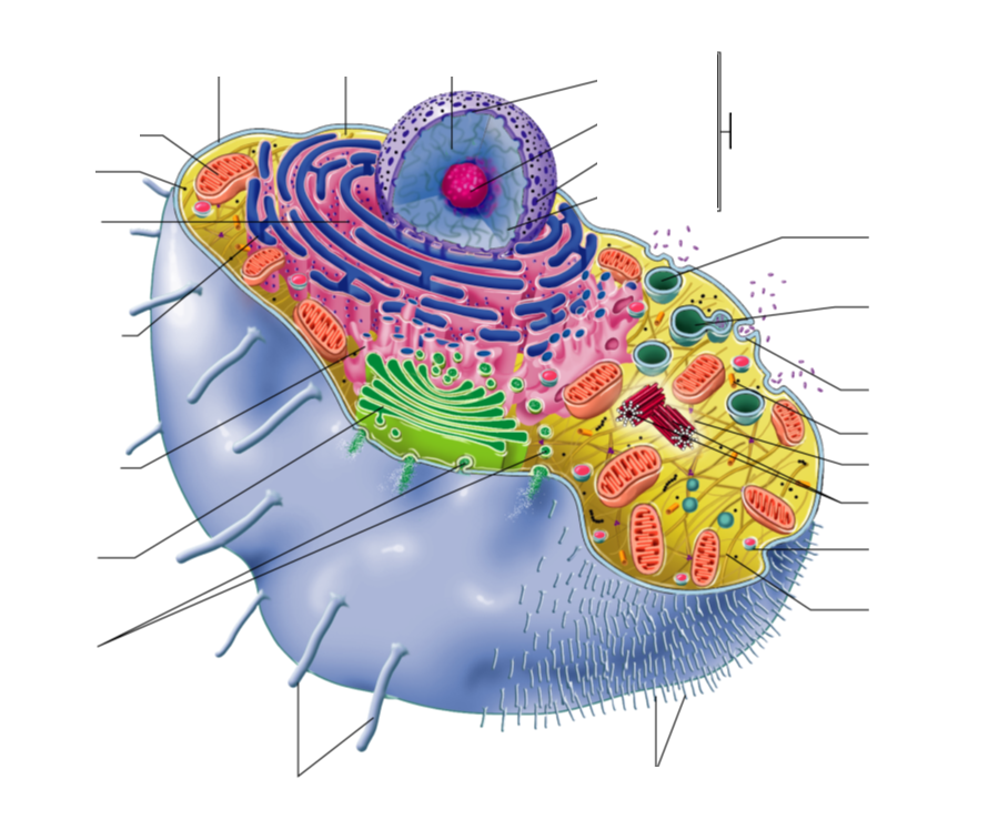
Diagram of a human cell with labels
Anatomy (Human Body) Labeling - Exploring Nature Arteries of the Lower Limb (Pelvis, Leg and Foot) Labeling. Arteries of the Upper Limb (Shoulder, Arm, Hand) Labeling. Blood Vessel Anatomy Labeling Thyroid gland: cells, tissues, labeled diagram (preview) - Human ... This is a sneak peek at the full tutorial about the thyroid gland histology. Watch the full video at Kenhub: , are you struggling with... Human Cell Diagram, Parts, Pictures, Structure and Functions Aug 09, 2018 · Parts of the Human Cell. The cell contains various structural components to allow it to maintain life which are known as organelles. All the organelles are suspended within a gelatinous matrix, the cytoplasm, which is contained within the cell membrane. One of the few cells in the human body that lacks almost all organelles are the red blood cells.
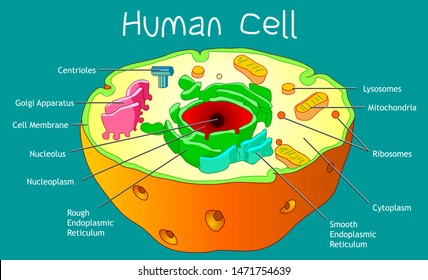
Diagram of a human cell with labels. A Labeled Diagram of the Animal Cell and its Organelles A Labeled Diagram of the Animal Cell and its Organelles There are two types of cells - Prokaryotic and Eucaryotic. Eukaryotic cells are larger, more complex, and have evolved more recently than prokaryotes. Where, prokaryotes are just bacteria and archaea, eukaryotes are literally everything else. Structure of Cell: Definition, Types, Diagram, Functions - Embibe What are the five cell structures? Ans: A cell consists of many different structures that have definite shapes, structures, and functions of their own. Some of these structures are (1) Cell Wall (2) Mitochondria (3) Chloroplast (4) Cell Membrane and (5) Nucleus . Q3. What is the structure of a human cell? Human Cells Printables and Diagrams - The Successful Homeschool These cells include: leukocytes, haematids, thrombocytes, ovum, sperm, sarcomeres, enterocytes, neurons, osteocytes, hepatocytes. They will learn the parts of a cell thanks to a labeled diagram. They will get to see what blood looks like under a microscope without needing to own a microscope. They get to color a cell and then label the parts. A Well-labelled Diagram Of Animal Cell With Explanation Well-Labelled Diagram of Animal Cell The Cell Organelles are membrane-bound, present within the cells. There are various organelles present within the cell and are classified into three categories based on the presence or absence of membrane. Listed below are the Cell Organelles of an animal cell along with their functions.
Cell Diagram | Free Cell Diagram Templates - Edrawsoft A free customizable cells diagram template is provided to download and print. Quickly get a head-start when creating your own cell diagram. Here is a simple cell diagram example created by Science Diagram Maker, which is available in different formats. Animal Cells: Labelled Diagram, Definitions, and Structure Animal Cells Organelles and Functions. A double layer that supports and protects the cell. Allows materials in and out. The control center of the cell. Nucleus contains majority of cell's the DNA. Popularly known as the "Powerhouse". Breaks down food to produce energy in the form of ATP. BYJUS BYJUS The following diagram shows cells of onion peel label class ... - Vedantu The following diagram shows human cheek cells, label the parts as observed by you. Answer. Verified. 106.2k+ views. Hint: The diagrams mentioned above are the internal structure of an onion peel and human cheek cells. In order to label them, we need to understand its anatomy and know about various structures present in it. Onion peel is an ...
Cell Membrane Diagram Labeled : Functions and Diagram Mar 22, 2021 · Cell Membrane Diagram. There are no organelles in the prokaryotic cells, i.e., they have no internal membrane systems. While lipids help to give membranes their flexibility, proteins monitor and maintain. We all keep in mind that the human body is very elaborate and a technique I found out to understand it is by means of the manner of human ... PDF Human Cell Diagram, Parts, Pictures, Structure and Functions Feb 06, 2017 · tail of the sperm cells which beats in a manner to allow the cell to move in a fluid medium. Functions of the Human Cell The functions of the human cell varies based on the type of cell and its location in the human body. All the organelles work together to keep the cell alive and allow it to carry out its specific function. Learn the parts of a cell with diagrams and cell quizzes Oct 28, 2021 · Two major regions can be found in a cell. The first is the cell nucleus, which houses DNA in the form of chromosomes. The second is the cytoplasm, a thick solution mainly comprised of water, salts, and proteins. The parts of a eukaryotic cell responsible for maintaining cell homeostasis, known as organelles, are located within the cytoplasm. diagram of the human labelling muscles body label key anatomy labeling labeled printable posterior. Label The Neuron Clip Art At Clker.com - Vector Clip Art Online . neuron diagram label unlabeled neurons nerve parts labeled anatomy clipart cliparts clip clker cells activity library vector name. Human Eye Diagrams With The Unlabeled
Human Cell Diagram Labeled - Diagram Media
03 Label the Cell Diagram | Quizlet Start studying 03 Label the Cell. Learn vocabulary, terms, and more with flashcards, games, and other study tools.
7 Best Images of Neuron Label Worksheet - Blank Neuron Cell Diagram, Synapse Neuron Worksheet ...
diagram human body label muscle labeling game muscular system label human labels quiz purposegames creative. Integumentary System Diagram To Label Lovely Hypodermis Skin Layers . integumentary anatomy. Digestive System | ClipArt ETC etc.usf.edu. system digestive clipart clip digestion diagram etc library unlabeled usf edu medium insertion codes

JQ Nursing Review: A&P Lecture 1: The Cell
Human Heart Diagram Labeled | Science Trends Daniel NelsonPRO INVESTOR. The human heart is an organ responsible for pumping blood through the body, moving the blood (which carries valuable oxygen) to all the tissues in the body. Without the heart, the tissues couldn't get the oxygen they need and would die. Along with lymphatic vessels, the blood, blood vessels, and lymph, the heart ...
muscular pictures 744×1179 | Anatomy System - Human Body Anatomy diagram and chart images
parts of a human cell | Diagram of the human cell illustrating the ... Apr 2, 2014 - parts of a human cell | Diagram of the human cell illustrating the different parts of the cell ... Pinterest. Today. Explore. ... This diagram of a human skeleton is labeled with 12 major bones, from skull to fibula. Nicole. Science. Human Cell Structure. Animal Cell Structure. Plant And Animal Cells.
Animal Cell Images, Stock Photos & Vectors | Shutterstock
Human Cell Organelles Labeling Diagram - Quizlet Human Cell Organelles Labeling STUDY Learn Flashcards Write Spell Test PLAY Match Gravity Created by Mackenna_Rios5 Terms in this set (8) Vesicles Transports molecules between organelles and the cell membrane Ribosome Makes Protein Mitochondria Makes ATP Smooth ER Makes lipids and vesicles Lysosomes

Educative diagrams: Digestive System Diagram
Diagram of human skin structure — Science Learning Hub Diagram of human skin structure. Add to collection. + Create new collection. Tweet. Rights: University of Waikato Published 1 February 2011 Size: 100 KB Referencing Hub media. The epidermis is a tough coating formed from overlapping layers of dead skin cells.

Questions And Answers On Labeled/Unlebled Diagrams Of A Human Cell / Question 14: Draw a labeled ...
Labeled Plant Cell With Diagrams - Science Trends The parts of a plant cell include the cell wall, the cell membrane, the cytoskeleton or cytoplasm, the nucleus, the Golgi body, the mitochondria, the peroxisome's, the vacuoles, ribosomes, and the endoplasmic reticulum. Parts Of A Plant Cell The Cell Wall Let's start from the outside and work our way inwards.

The diagram shows a certain kind of cell with all of its major parts labeled. Which statement is ...
by pulpbits.com | Human cell diagram, Cell diagram, Animal cell drawing Plant Cell Science Diagram Clipart Set includes: Three diagrams (one labeled, one with blank labels and diagram alone) plus 9 mini-diagrams of different "cell parts." Each image comes in color and line art. Elements are high resolution 300 dpi png format with transparent backgrounds.
Images 01. Introduction and Terminology | Basic Human Anatomy
Label Diagram Human Body Stock Illustrations - Dreamstime Download 188 Label Diagram Human Body Stock Illustrations, Vectors & Clipart for FREE or amazingly low rates! New users enjoy 60% OFF. 187,764,331 stock photos online. ... Labeled diagram of the neuron. Nerve cell that is the main part of the nervous system. The circulatory system. The circulatory or cardiovascular human body system medical ...

Cell Review Guide Answers in 2020 | Human cell structure, Human cell diagram, Animal cell structure
Labeled Diagram of the Human Lungs - Bodytomy Given below is a labeled diagram of the human lungs followed by a brief account of the different parts of the lungs and their functions. Each lung is enclosed inside a sac called pleura, which is a double-membrane structure formed by a smooth membrane called serous membrane.

An Annotated Diagram Of A Human Cell
Cell: Structure and Functions (With Diagram) - Biology Discussion Eukaryotic Cells: 1. Eukaryotes are sophisticated cells with a well defined nucleus and cell organelles. 2. The cells are comparatively larger in size (10-100 μm). 3. Unicellular to multicellular in nature and evolved ~1 billion years ago. 4. The cell membrane is semipermeable and flexible. 5. These cells reproduce both asexually and sexually.

Ciencias 4to grado: Human Body- Tissues
Human Cell Diagram, Parts, Pictures, Structure and Functions Aug 09, 2018 · Parts of the Human Cell. The cell contains various structural components to allow it to maintain life which are known as organelles. All the organelles are suspended within a gelatinous matrix, the cytoplasm, which is contained within the cell membrane. One of the few cells in the human body that lacks almost all organelles are the red blood cells.

Animal Cell Model: What To Use - YouTube
Thyroid gland: cells, tissues, labeled diagram (preview) - Human ... This is a sneak peek at the full tutorial about the thyroid gland histology. Watch the full video at Kenhub: , are you struggling with...

The Human Egg Cell Explained For Egg Donors - Altrui Egg Donation Agency
Anatomy (Human Body) Labeling - Exploring Nature Arteries of the Lower Limb (Pelvis, Leg and Foot) Labeling. Arteries of the Upper Limb (Shoulder, Arm, Hand) Labeling. Blood Vessel Anatomy Labeling

Biology Unit: Cells, Organs, Systems - Ms. Corner Gardiner's Classes
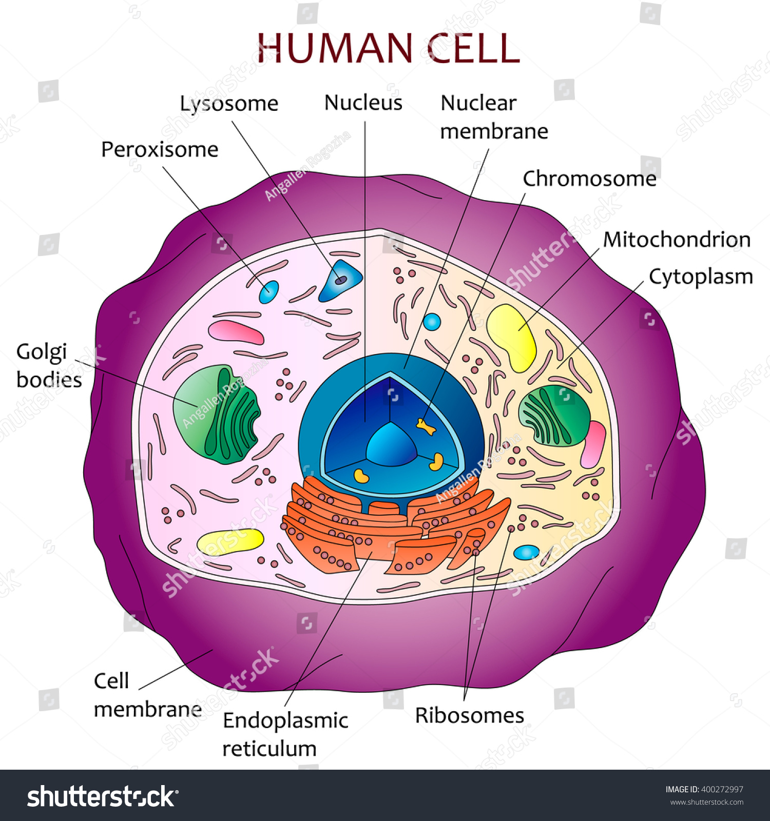
Human Cell Diagram Stock Vector 400272997 - Shutterstock
Questions And Answers On Labeled/Unlebled Diagrams Of A Human Cell : Plant Cell Definition ...
Post a Comment for "45 diagram of a human cell with labels"